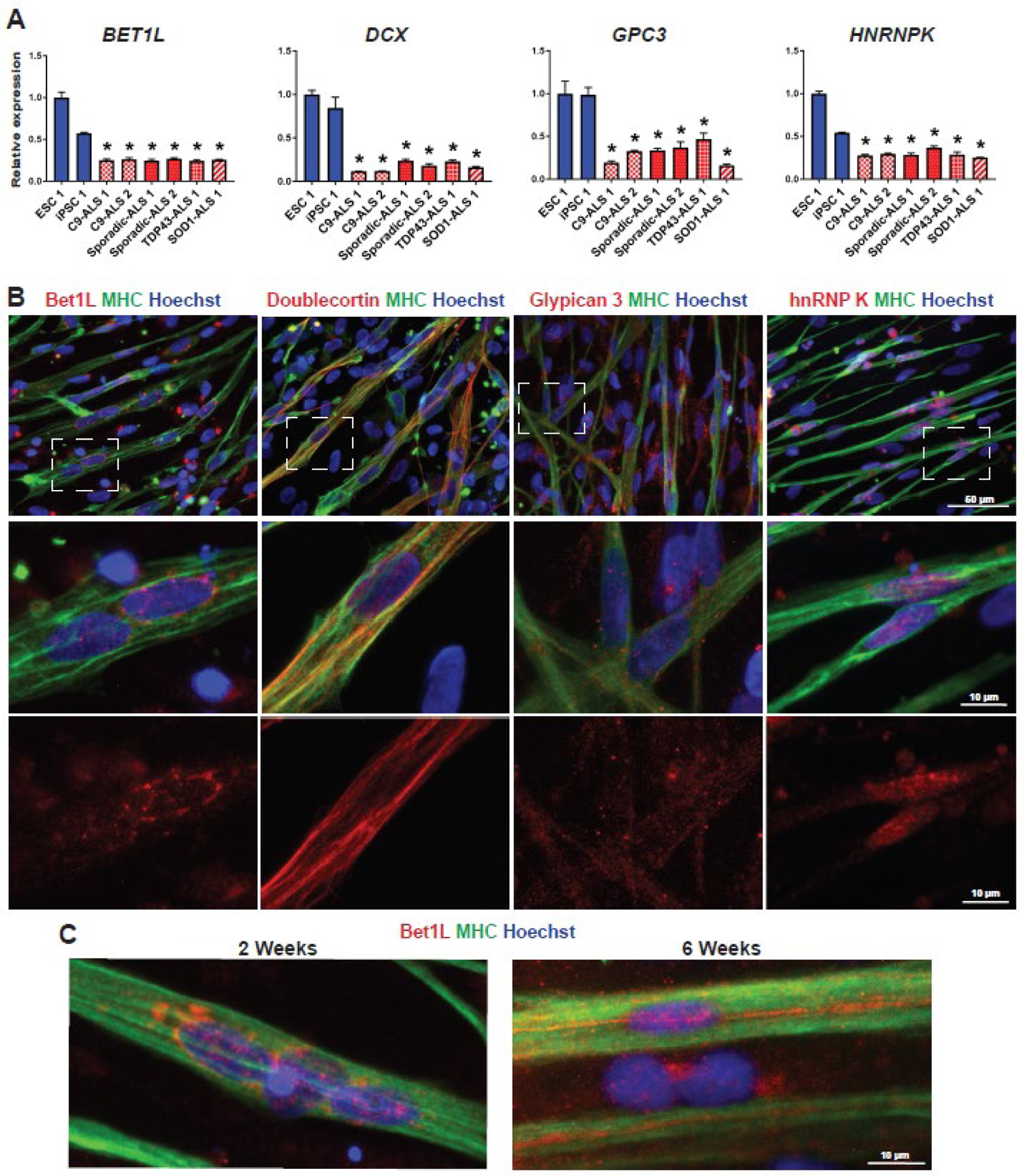
Figure 2. Expression of the four proteins of interest in iPSC-derived skeletal myocytes.

(A) RT-qPCR of day 14 skeletal myocytes was used to confirm down-regulation of each of the four genes identified from RNA sequencing. Data was normalized to control line ESC 1 and analyzed using one-way ANOVA followed by Tukey’s post-hoc multiple comparisons test. *P<0.01 compared to ESC 1 and iPSC 1 (B) Representative images of each protein of interest in healthy control myocytes at day 14. Skeletal myocytes are indicated by positive expression of myosin heavy chain (MHC). The middle row is enlarged from the dashed squares, and the bottom row shows red channel only. (C) The localization of Bet1L protein changed over time, with a more aligned expression pattern in mature myocytes around 6 weeks.
