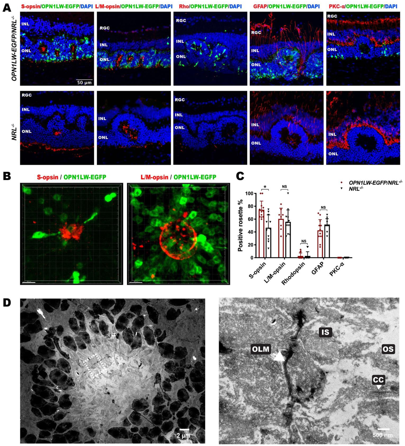
Fig. 3. Cellular composition of rosettes in OPN1LW-EGFP/NRL−/− mice.

(A) IHC images of P35 OPN1LW-EGFP/NRL−/− retinal sections stained with retinal specific markers, including S-opsin, L/M-opsin, Rhodopsin, GFAP, and PKC-α, with age-matched NRL−/− retina as control.
(B) Three-dimensional reconstitution of S-opsin+ and L/M-opsin+ rosette on retina flat mount of OPN1LW-EGFP/NRL−/− mice (P35).
(C) Quantification of rosettes that were positive for each marker in OPN1LW-EGFP/NRL−/− and NRL−/− mice. * p < 0.0001. NS: not significant.
(D) Transmission electron microscopy of a representative rosette in OPN1LW-EGFP/NRL−/− mice (P35). The magnified image is on the right side.
Abbreviations: RGC: retinal ganglion cell; INL: inner nuclear layer; ONL: outer nuclear layer; OLM: outer limiting membrane; IS: inner segment; CC: connecting cilium; OS: outer segment.
