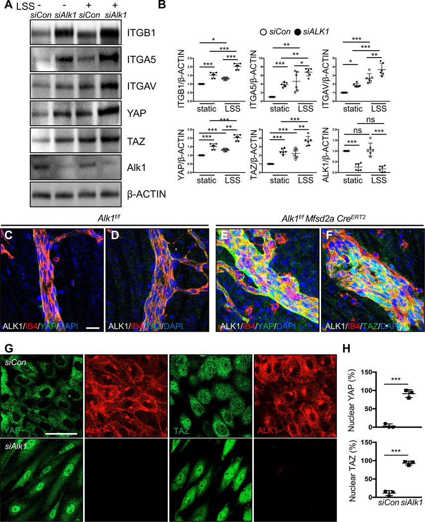
Figure 6. ALK1 controls YAP/TAZ expression and localization.

(A) Western blot analysis of HUVECs transfected with control and ALK1 siRNAs followed by 18 h exposure to LSS (15 dynes/cm2). (B) Quantification of ITGB1, ITGA5, ITGAV, YAP or TAZ levels normalized to β-ACTIN. *P<0.05, **P<0.01, ***P<0.001, Two-way ANOVA with Sidak’s multiple comparison test. (C-F) YAP and TAZ (green), ALK1 (gray), IB4 (red), DAPI (blue) staining of retinal flat mounts from P8 Alk1f/f (C-D) or Alk1f/f Mfsd2aCreERT2 (E-F) pups. A scale bar: 20 μm (C-F) (G) YAP or TAZ (green) and ALK1 (red) staining of siCon and siALK1 HUVECs. A scale bar: 50 μm. (H) Quantification of nuclear YAP and TAZ from siCon and siALK1 transfected HUVECs. ***P<0.001, n = 3 independent experiments. Two-tailed unpaired t-test between siCon and siALK1.
