
Figure 7. YAP/TAZ inhibition improves AVM formation in Alk1 mutant retinas.
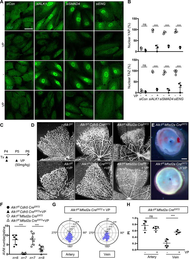
(A) YAP and TAZ staining of siCon, ALK1, SMAD4 or ENG siRNAs transfected HUVECs treated with DMSO or Verteporfin (VP, 5 μM) for 6 h. Nuclear YAP/TAZ localization in siALK1, siSMAD4 or siENG ECs is blocked by VP treatment. A scale bar: 50 μm. (B) Quantification of nuclear YAP and TAZ from siCon, siALK1,siSMAD4 and siENG transfected HUVECs. ***P<0.001, ns: nonsignificant, Two-way ANOVA with Tukey’s multiple comparisons test. (C) Experimental strategy to assess the effects of YAP/TAZ inhibition in EC specific Alk1 deleted vasculature. Arrowheads indicate the time course of Tx (100 μg) and VP (50mg/kg) or vehicle administration. (D) IB4 staining of P6 retinal flat mounts from VP injected Alk1f/f, Alk1f/f CDH5 CreERT2 or Alk1f/f Mfsd2a CreERT2 mice. (E) Stereomicroscopy images of vehicle or VP injected Alk1f/f Mfsd2a CreERT2 retinas. (F) Quantification of the AVM number/retina. Each dot represents one retina. n = 6–8 retinas per group. Error bars: SEM. ***P-value < 0.001, One-way ANOVA with Sidak’s multiple comparisons test. (G) Angular histograms showing polarization angles of artery and vein from Alk1f/f Mfsd2a CreERT2 with VP. (H) PI box plots of Alk1f/f Mfsd2a CreERT2 with vehicle or VP. n=5–6 retinas, Error bars: SEM, ***P-value < 0.001, ns: nonsignificant, Two-way ANOVA with Tukey’s multiple comparisons test. Scale bars : 50 μm (A), 500 μm (D), 300 μm (E)