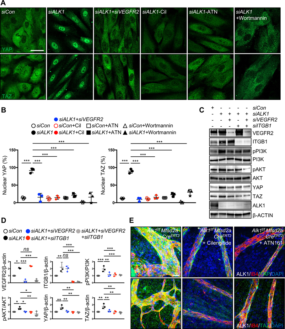
Figure 8. VEGFR2, integrin and PI3K function upstream of YAP/TAZ in Alk1 mutants.

(A) YAP and TAZ staining for siCon, siALK1 and siALK1+siVEGFR2 transfected HUVECs and ALK1 siRNA transfected HUVECs treated with cilengitide (Cil, 5 μM), ATN161 (ATN, 5 μM) and wortmannin (100 mM) for 12 h. Nuclear YAP/TAZ localization in siALK1 ECs is blocked by siVEGFR2, cilengitide, ATN161 and wortmannin treatment. (B) Quantification of nuclear YAP and TAZ from for siCon, siALK1 and siALK1+siVEGFR2 transfected HUVECs and ALK1 siRNA transfected HUVECs treated with cilengitide, ATN16, wortmannin. ***P<0.001, n = 3 independent experiments. Error bars: SEM. ***P-value < 0.001, Two-way ANOVA with Tukey’s multiple comparisons test. (C) Western blot analysis of HUVECs transfected with control, ALK1, ALK1+VEGFR2, ALK1+ITGB1 or ALK1+VEGFR2+ITGB1 siRNAs. (D) Quantification of pPI3K/PI3K, pAKT/AKT and YAP or TAZ levels normalized to β-ACTIN. *P<0.05, **P<0.01, ***P<0.001, ns: nonsignificant, One-way ANOVA with Sidak’s multiple comparisons test. (E) YAP and TAZ (green), ALK1 (white), IB4 (red) and DAPI (blue) staining of retinal flat mounts from cilengitide or ATN161 injected Alk1f/f Mfsd2a CreERT2 P6 mice. Scale bars: 50 μm (A), 20 μm (E)
