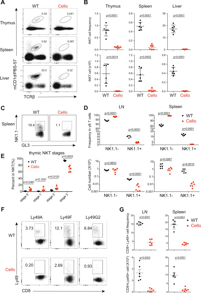
Fig. 3.
Cellophane mice have decreased numbers of thymic DP cells and lack iNKT cells, NK1.1+ γδ T cells and Ly49+ CD8 cells. a Flow cytometry analysis of CD1d-tet+ iNKT cells (black gate) from thymus, spleen, and liver in wild-type (WT) and Cellophane (Cello) homozygous mice as indicated. b Percentages and absolute numbers of iNKT cells (mCD1d/PBS-57+ TCRβ+) in the thymus, spleen, and liver. c Flow cytometry analysis of NK1.1− and NK1.1+ γδ T cells (black square gates) from the spleen of WT and Cellophane mice. d Percentages within total γδ T cells and absolute numbers of NK1.1− and NK1.1+ γδ T cells in spleen and LN. e Percentage within total thymic iNKT cells of cells from stages 0–3, as defined by CD24, CD44, and NK1.1 expression. f Flow cytometry analysis of CD8+ CD44+ Ly49A+/Ly49F+/Ly49G2+ T cells (black square gates) from LN and spleen from WT and Cellophane mice. g Percentage within CD8+CD44+ T cells and absolute number of CD8+ CD44+ Ly49+ T cells in spleen and LN. Each dot represents an individual mouse. Data are representative of two to four independent experiments with six mice in total in each panel. The statistical analysis was performed using an unpaired Student’s t test

