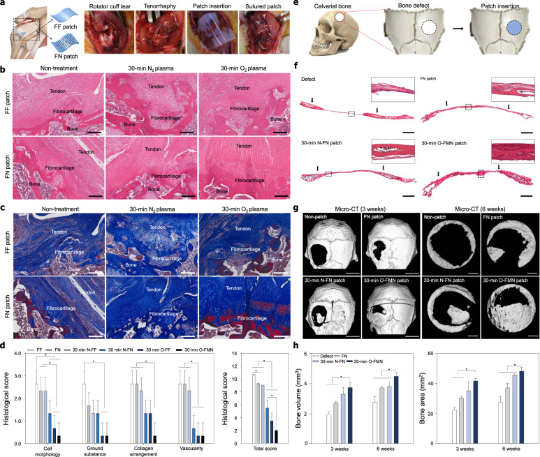
Fig. 4. Effect of the O-FMN patches on the tendon and bone tissue regeneration.
a Surgical procedure for RC tendon repair. All patches were grafted onto the defected tendon tissue after tenorrhaphy of the torn RC tendon (n = 3 for each group). b Representative histologic images of H&E staining and c Masson trichrome staining of the insertion site of FF, FN, 30-min N-FF, 30-min N-FN, 30-min O-FF, and 30-min O-FMN patches onto the supraspinatus tendon 4 weeks after repair. Scale bars = 200 µm. d Semiquantitative analysis of the histological evaluation scores on repaired tendon tissues of RC tear animal models. e Surgical procedure for rat calvarial bone repair (n = 5 for each group). f Representative histologic images of H&E staining of the insertion site of FN, N-FN, and O-FMN patches onto rat calvarial bone 6 weeks after repair. Scale bars = 1 mm. g Representative micro-CT image after 3 and 6 weeks of repair. Scale bars = 5 mm (3 weeks). Scale bars = 1 mm (6 weeks). h Quantitative analysis of bone volume and area of the bone regeneration site after 3 and 6 weeks of repair. Error bars = mean ± standard deviation (*P < 0.05).

