Figure 5.
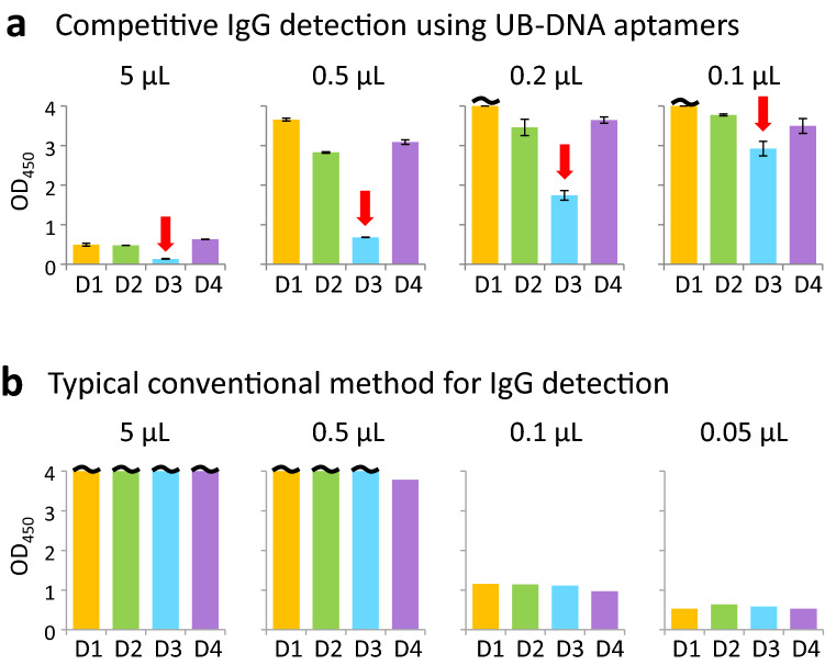
Comparison of the serotype-specific IgG detection by the competitive and conventional ELISA formats using a clinical sample (plasma, PD2-4). (a) The competitive Apt/Ab ELISA format (Fig. 1c), displaying the inhibition of the aptamer–DEN-NS1 binding by IgGs in our competitive Apt/Ab ELISA format using 0.1 to 5 µL of a clinical sample obtained within 4 days after fever onset. The amounts of the recombinant DEN-NS1 (The Native Antigen Company) added to each well (50 μL) were 350 pg for DEN1-NS1, 350 pg for DEN2-NS1, 450 pg for DEN3-NS1, and 200 pg for DEN4-NS1. Red arrows indicate the higher inhibition of the spiked DEN-NS1 detection among the four serotypes of DEN-NS1, suggesting that the IgG in the sample is more specific to DEN3-NS1. The results revealed that the current infection of PD2-4 might be secondary, and the past infection might be DENV serotype 3, although the Panbio LFA kit did not detect IgGs from the sample (Supplementary Table 2). (b) Direct IgG detection by a typical conventional ELISA method (Fig. 1d) using 0.05–5 μL of the sample. Each well was coated with 50 ng recombinant DEN-NS1 protein.

