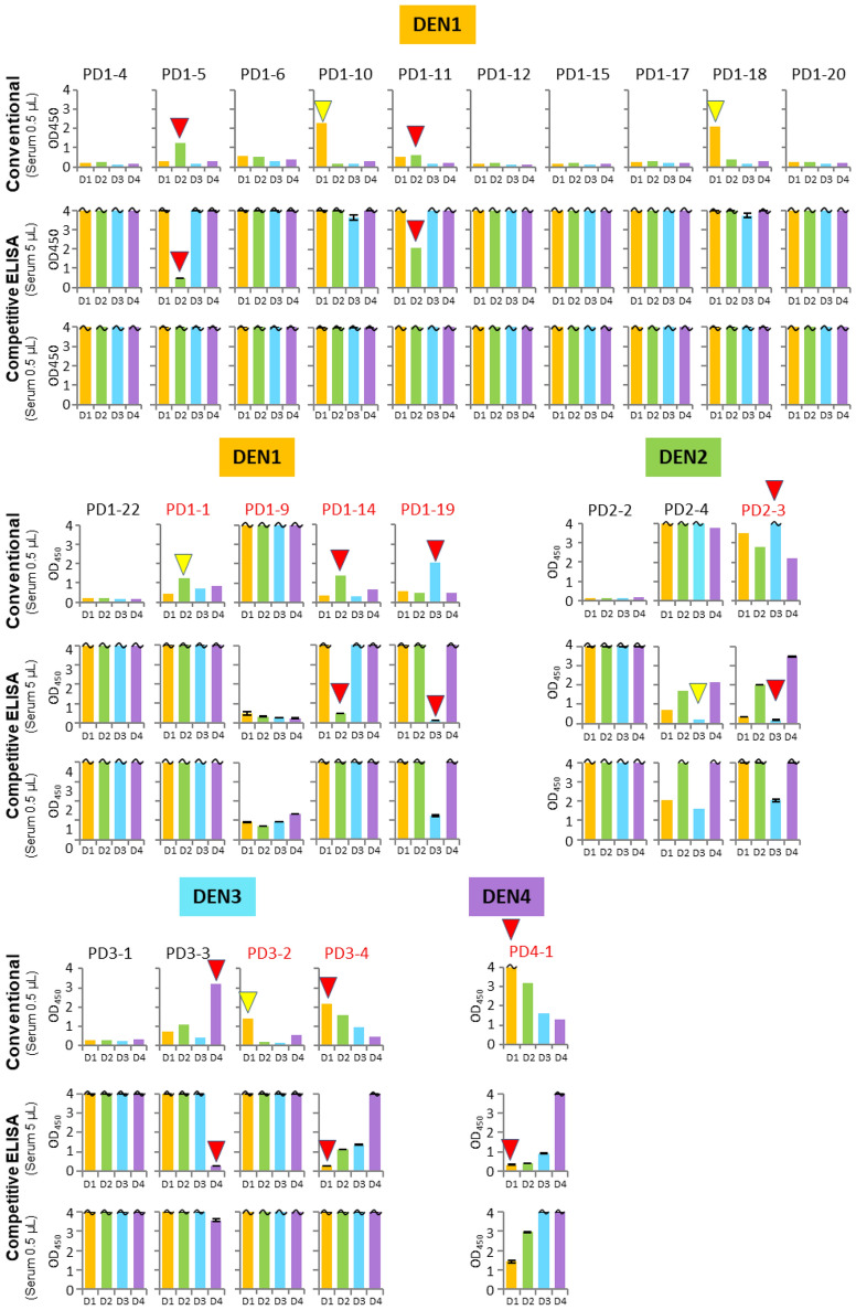
Figure 6.
Comparison of the IgG detection sensitivity and selectivity using various clinical samples with two types of ELISA. Conventional: the direct IgG detection by the typical ELISA method in Figs. 1d and 5b using 0.5 µL of the serum or plasma samples. Competitive ELISA: the aptamer–DEN-NS1 binding inhibition by our competitive Apt/Ab ELISA (Figs. 1c and 5a) using 0.5 µL and 5 µL of the serum or plasma samples. The amounts of the spiked DEN-NS1 are indicated in Supplementary Fig. 1. The similar patterns of the serotype-specific IgG detection by both ELISA formats are indicated by red triangles. IgG detection patterns that differ between the conventional and competitive ELISA formats are indicated by yellow triangles. The patient sample names (indicated in red) were assigned as secondary infection by the LFA test.

