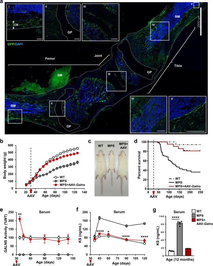
Fig. 1. AAV9-Galns treatment corrects MPSIVA pathology.
a Widespread transduction of femur and tibia after intravascular administration of 6.67 × 10¹³ vg/kg AAV9-GFP vectors to 4-weeks-old wild-type (WT) rats. Two weeks later, GFP signal was detected in compact bone (CB) (I), areas surrounding growth plate (GP) (II), joint (III), trabecular bone (IV), and endosteum (V). n = 3 animals/group. Scale bars: 2000 µm; insets, 300 µm. BM bone marrow, SM skeletal muscle. Afterward, 4-weeks-old MPSIVA male rats were treated with 6.67 × 10¹³ vg/kg AAV9-Galns vectors. b Body weight of WT (n = 21), MPSIVA untreated (n = 26), and AAV9-Galns-treated (n = 20) rats. c Representative image of 2-months-old WT, untreated and AAV9-Galns-treated MPSIVA rats. d Kaplan–Meier survival analysis, WT (n = 17), untreated (n = 76), and AAV9-Galns-treated MPSIVA (n = 32) rats. e Circulating GALNS activity of WT (n = 5), untreated (n = 5) and AAV9-Galns-treated MPSIVA (n = 18) rats. WT activity was set to 100%, which ranged from 94 to 133 nmol/17 h/mg. f, g Serum KS levels at different ages of WT (n = 4), untreated (n = 5) and AAV9-Galns-treated MPSIVA (n = 7) rats (f) and of 1-year-old WT (n = 3), untreated (n = 4) and AAV9-Galns-treated MPSIVA (n = 2) rats (g). All results are shown as mean ± SEM and P-values of one-way Anova test are indicated (95% confidence interval). *P < 0.05, **P < 0.01, and ****P < 0.0001 vs. untreated MPSIVA rats. Source data are provided as a Source Data file.

