Fig. 4.
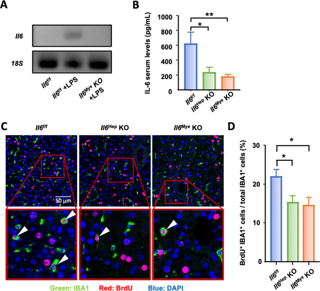
Hepatocytes and KCs are important sources of IL-6 after PHx. A RT-qPCR confirming Il6 deletion in KCs from Il6Mye-KO mice. Il6 mRNA expression in KCs isolated from control Il6f/f mice injected with PBS or LPS and Il6Mye-KO mice injected with LPS (3 h injection). B Serum IL-6 levels in Il6f/f, Il6Hep-KO, and Il6Mye-KO mice 3 h after PHx (n = 6/group). C, D Immunofluorescence analysis of liver tissue sections from Il6f/f, Il6Hep-KO, and Il6Mye-KO mice collected 48 h after PHx (n = 6/group) and stained with anti-IBA1 (green) and BrdU (red) antibodies. BrdU was injected 2 h before sacrifice, as shown in panel (C). Arrowheads represent proliferating KCs. The quantification of macrophage proliferation in the liver 48 h after PHx is shown in panel (D). The values are expressed as the mean ± SEM. *P < 0.05, **P < 0.01

