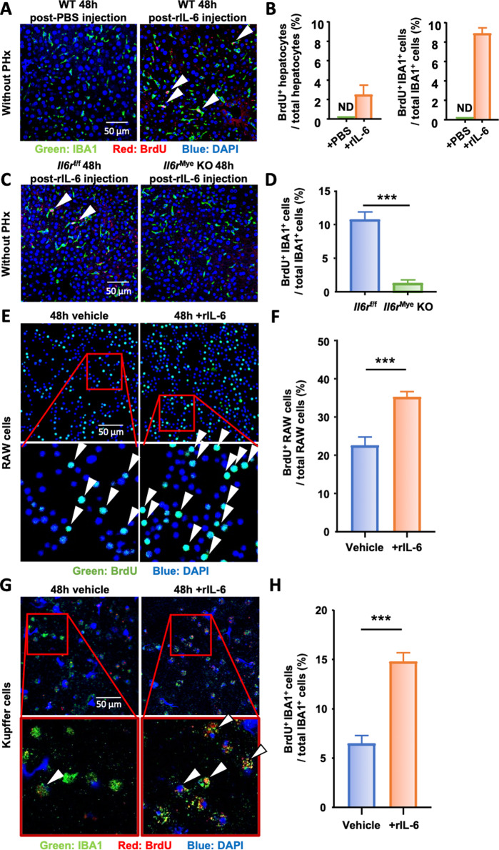
Fig. 6.
IL-6 stimulates macrophage proliferation in vivo and in vitro. A, B Naïve wild-type mice (without PHx) were intravenously injected with PBS (control) or rIL-6, and liver tissues were collected 48 h after injection (n = 7/group) and subjected to immunofluorescence staining with antibodies against IBA1 (green) and BrdU (red). Arrowheads represent proliferating KC. The quantifications of BrdU+ hepatocytes and BrdU+ IBA1+ cells are shown in panel B. ND: not detected. C, D Il6rf/f and Il6rMye-KO mice without PHx were intravenously injected with rIL-6 (n = 7/group), and liver tissues were collected 48 h after injection and subjected to immunofluorescence staining with antibodies against IBA1 (green) and BrdU (red). Arrowheads represent proliferating KC. The quantification of proliferating KCs in livers from Il6rf/f and Il6rMye-KO mice 48 h after intravenous injection of rIL-6 is shown in panel (D). E, F Immunofluorescence staining of RAW cells 48 h after exposure to rIL-6 or control medium (vehicle) with antibodies against BrdU (green). Proliferating RAW cells are identified by arrowheads. The quantification of proliferating RAW cells is shown in panel (F). G, H Immunofluorescence staining of freshly isolated KCs 48 h after exposure to rIL-6 or control medium (vehicle) with antibodies against IBA1 (green) and BrdU (red). The arrowheads represent proliferating KC. The quantification of KC proliferation is shown in panel (H). BrdU was injected 2 h before sacrifice in panels (A) and (C) and was added to the culture medium 2 h before collecting the cells in panels (E) and (G). The values are expressed as the mean ± SEM. ***P < 0.001

