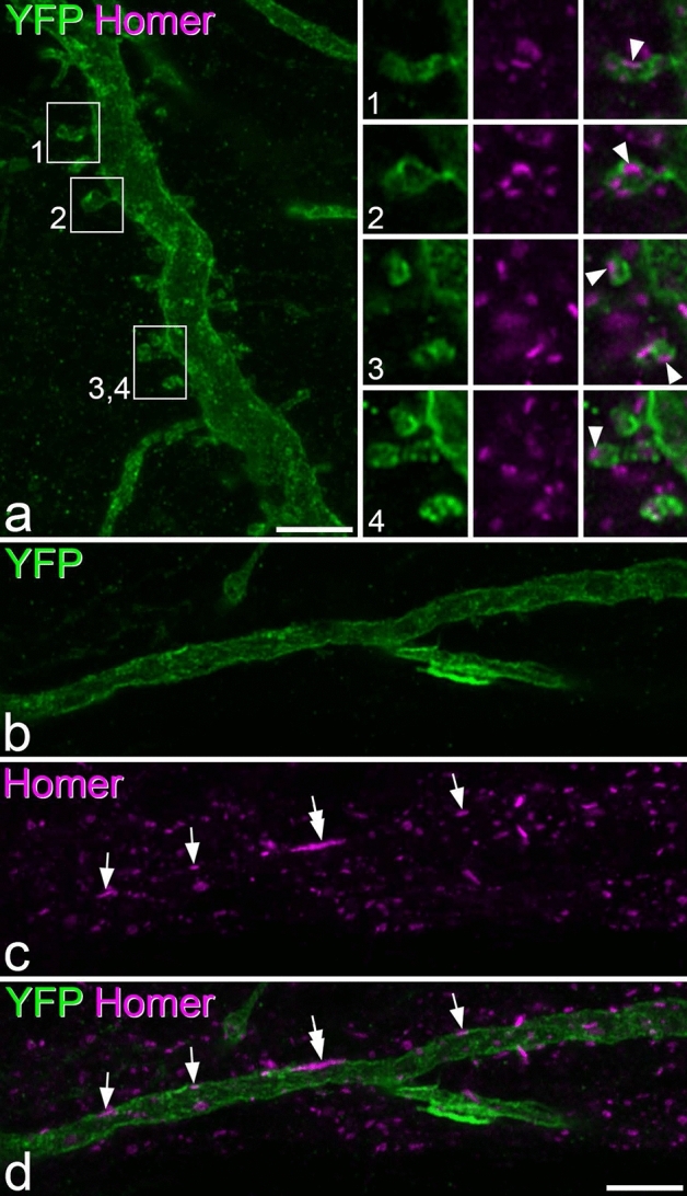
Figure 11.

Homer puncta on the dendritic shafts and spines of YFP-positive lamina I cells in Phox2a::Cre;Rosa26LSL- ChR2-EYFP mice. (a) A region of dendrite from Cell 3 (shown in Fig. 9) that has numerous dendritic spines. The insets show higher magnification views of these spines. The tissue has been reacted to reveal YFP (green) and Homer (magenta). Homer puncta can be seen in each of the dendritic spines (arrowheads) in the insets. (b–d) Part of a dendritic shaft from the same cell. This region has few dendritic spines, but there are several Homer puncta in the membrane (arrows). One of these (double arrow) is particularly large (approximately 3 μm long). Confocal images are projections of 11 optical sections (main part of (a) and 4 optical sections (b–d) all at 0.3 μm z-separation. Insets in (a) are all single optical sections. Scale bars: (a,b–d) = 5 μm.
