Figure 6.
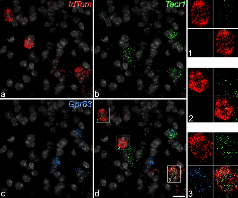
Fluorescence in situ hybridisation with RNAscope in a horizontal section from a Phox2a::Cre;Rosa26LSL-tdTomato mouse. The section was reacted with probes for tdTom (red), Tacr1 (green) and Gpr83 (blue) mRNAs, and these are shown separately in (a–c) and combined in (d). The nuclear counterstain NucBlue is shown in grey. Three tdTom-positive cells are present (numbered 1–3 in (d)). These are illustrated at higher magnification in the insets, and in each of these the top row shows labelling for tdTom and Tacr1 and the bottom row labelling for Gpr83 together with a merged image. All 3 cells are positive for Tacr1 (based on the presence of more than 5 transcripts), while cell 3 is positive for Gpr83. Note that there are sparse scattered transcripts for tdTom, which are also seen in Rosa26LSL-tdTomato mice, and presumably result from a low-level “leak” of tdTomato expression. However, these can easily be distinguished from the labelling in the putative Phox2a-positive lamina I neurons. Images are projections of confocal optical sections (1 μm z-separation) through the full thickness of the section. Scale = 20 μm.

