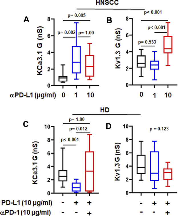
FIGURE 3.

αPD-L1 treatment increases K+ channel activity in HNSCC patients. (A) KCa3.1 and (B) Kv1.3 conductance values (G) measured with and without the αPD-L1 antibody, atezolizumab (1 and 10 μg/ml for 6 h) in activated CD8+ PBTs of HNSCC patients. (C) KCa3.1 and (D) Kv1.3 G measured in presence of PD-L1 and αPD-1 antibody pembrolizumab in activated CD8+ PBTs of HDs. Activated cells were treated with plate-bound PD-L1 (PD-L1-Fc 10 μg/ml) +/- αPD-1 (untreated cells were used as a control) and activated for 72 h using PMA/Ionomycin. αPD-1 was added to treatment group for 6 h. Data in the lower and upper bound of the box represent 25th and 75th percentiles respectively. Median values are shown as horizontal lines. The lower and upper error bars represents 10th and 90th percentile respectively, n = 8–23 cells from 3 HNSCC patients, n = 30 cells from 6 HDs (control and PD-L1) and n = 15 cells from 3 HDs (PD-L1 + αPD-1). Five cells were recorded for each individual donor. Data in (A,C,D) were analyzed by ANOVA on ranks test (p < 0.001) followed by Dunn’s post hoc analysis. Data in (B) were analyzed by One way ANOVA (p < 0.001) followed by Holm-Sidak test.
