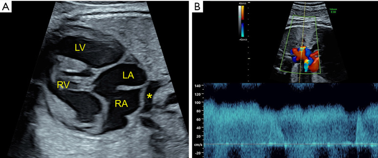
Figure 5.
Total anomalous pulmonary venous connection in a fetus with heterotaxy, atrioventricular canal defect, and pulmonary stenosis. (A) The pulmonary venous confluence (*) is seen behind the right (RA) and left atrium (LA). (B) Pulse wave Doppler of the vertical vein showing non-phasic high velocity flow, consistent with obstruction, as it enters the innominate vein. RV, right ventricle; LV, left ventricle.

