Figure 8.
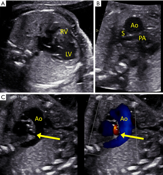
Fetus with pulmonary atresia and intact ventricular septum. (A) The right ventricle (RV) is severely hypoplastic and hypertrophied compared to the dilated left ventricle (LV). (B) Abnormal three-vessel view demonstrating a dilated aorta (Ao) and small pulmonary artery (PA). (C) Reverse oriented (arrow) ductus arteriosus with aorta to pulmonary artery flow predicting neonatal ductal dependence for pulmonary blood flow. S, superior vena cava.

