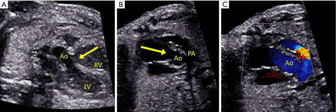
Figure 9.
Fetus with non-ductal dependent tetralogy of Fallot. (A) Outflow tract imaging showing the overriding aorta (Ao) and ventricular septal defect (arrow). (B) Short axis view of the aorta and anterior malalignment ventricular septal defect (arrow), with the pulmonary artery (PA) smaller than the aorta. (C) Color flow shows mild flow acceleration across the mildly hypoplastic pulmonary valve.

