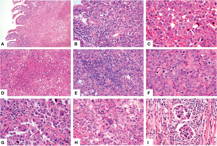
Figure 3.
Histologic features of the SMARCA4-deficient undifferentiated carcinoma. The neoplastic cells involved the mucosa [(A), X100]. Higher magnification showed several glands [(B), X400], noncohesive rhabdoid cells [(C), X400], multiple necrosis [(D), X200], patchy lymphocytes and plasma cells infiltration [(E), X400], obvious mitotic figures [(F), X400], multinucleated tumor cells [(G), X400] and large cells [(H), X400]. Multiple lymphovascular permeation was present [(I), X400].

