Figure 4.
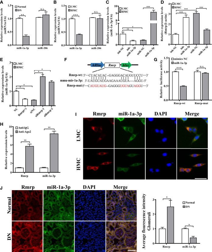
Rmrp functioned as a ceRNA by sponging miR-1a-3p. (A) The expression of miR-1a-3p and miR-206 were detected by qRT-PCR in the renal tissues of db/db DN mice and normal mice (n = 5/group). (B) The expression of miR-1a-3p and miR-206 were detected by qRT-PCR in LMC and HMC. (C) The expression levels of miR-1a-3p were detected by qRT-PCR after transfection with miR-1a-3p mimics and inhibitor in MCs. (D) The expression of Rmrp regulated by miR-1a-3p mimics and inhibitor were detected by qRT-PCR. (E) The expression of miR-1a-3p were detected in LMCs transfected with Rmrp (+) and in HMC transfected with siRmrp. (F) Schematic illustration revealed the base complementation of miR-1a-3p with Rmrp and mutant (Mut) sequences. (G) Luciferase assay was used to test relative luciferase activities of Rmrp in MC co-transfected with the indicated miR-1a-3p or control vector. (H) Anti-AGO2 RIP was performed in HMC and the RNA levels of Rmrp and miR-1a-3p were determined by qRT-PCR. (I) The co-localization of Rmrp and miR-1a-3p was observed in LMC and HMC (Scale bar, 50 μm) by FISH assay. (J) The co-localization of Rmrp and miR-1a-3p was observed in the renal tissues of db/db DN mice and normal mice by FISH assay and quantitative analysis (Scale bar, 50 μm). Data were represented as the mean ± SD of three independent experiments; *p < 0.05, **p < 0.01, and ***p < 0.001. ns, no significant.

