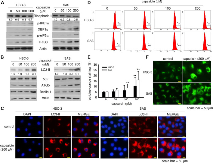
FIGURE 2.
Capsaicin dose-dependently down-regulates ribophorin II and enhances autophagy in HSC-3 and SAS cells. (A,B) Cells were treated with different concentrations of capsaicin for 48 h and the levels of ER stress- and autophagy-related proteins were measured by Western blot analysis. (C) Cells were treated with or without 200 μM capsaicin for 48 h followed by immunofluorescence staining to detect the levels of LC3-II. (D,E) Cells were treated with different concentrations of capsaicin for 48 h. The cells were then collected in microcentrifuge tubes and treated with 0.1 μg/ml acridine orange for 30 min before being analyzed by flow cytometry. (F) After treatment with capsaicin, cells were stained with the DAPGreen reagent, as described in the Materials and Methods. Data are representative of five to ten independent experiments. Scale bar = 50 μm.

