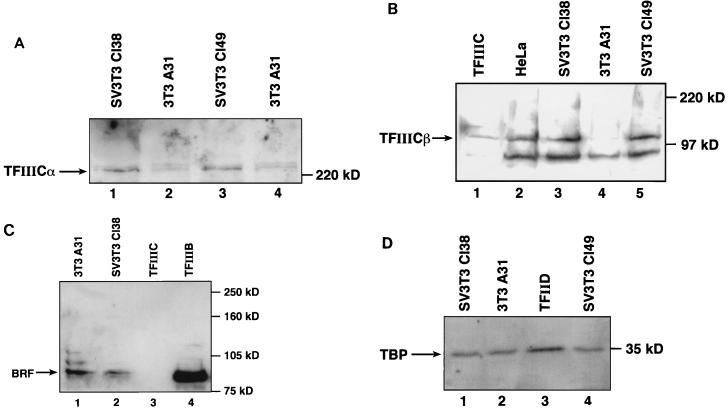
FIG. 1.
The SV40-transformed cell lines Cl38 and Cl49 express elevated levels of TFIIIC2 subunits. (A) Whole-cell extracts (57 μg of protein) prepared from Cl38 (lane 1), A31 (lanes 2 and 4), and Cl49 (lane 3) cells were resolved on an SDS–7.8% polyacrylamide gel and then analyzed by Western immunoblotting with anti-TFIIICα antibody Ab4. Lanes 2 and 4 show extracts prepared from two separate batches of A31 cells. (B) CHep-1.0 fraction (15 μg of protein) containing TFIIIC2 (lane 1) and whole-cell extracts (38 μg of protein) prepared from HeLa (lane 2), Cl38 (lane 3), A31 (lane 4), and Cl49 (lane 5) cells were resolved on an SDS–7.8% polyacrylamide gel and then analyzed by Western immunoblotting with anti-TFIIICβ monoclonal antibody clone 46. (C) Whole-cell extracts (50 μg of protein) prepared from A31 (lane 1) and Cl38 (lane 2) cells, CHep-1.0 (12 μg of protein) TFIIIC2 fraction (lane 3), and A25(0.15) (1.6 μg of protein) TFIIIB fraction (lane 4) were resolved on an SDS–7.8% polyacrylamide gel and then analyzed by Western immunoblotting with anti-BRF antibody 330. (D) Whole-cell extracts (50 μg of protein) prepared from Cl38 (lane 1), A31 (lane 2), and Cl49 (lane 4) cells and PC-D (5.6 μg of protein) TFIID fraction (lane 3) were resolved on an SDS–7.8% polyacrylamide gel and then analyzed by Western immunoblotting with anti-TBP antibody SL30.

