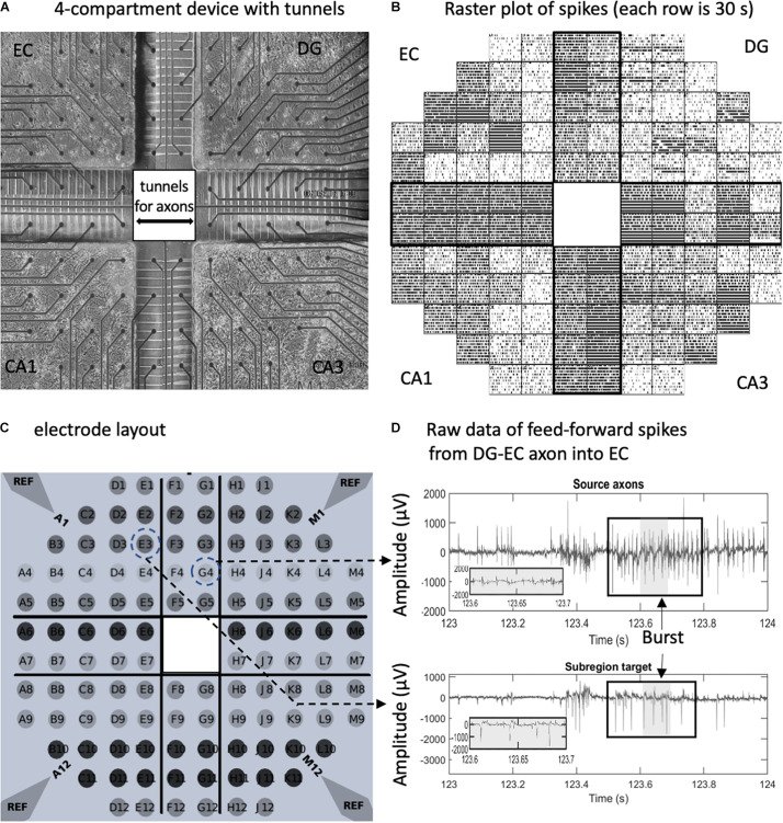
FIGURE 1.
In vitro neuron culture of four separate hippocampal subregions with axonal interconnections aligned on a 120-electrode array. (A) Phase contrast image after 3 weeks in culture. Spacing between the 30-μm diameter electrodes is 0.2 mm. (B) Raster plot of spikes detected on individual electrodes over 5 min (30 s/row). Each tick is one spike. (C) Electrode array layout. Reference ground electrodes are seen in the corners. (D) Electrical feedback signals from the indicated electrodes with source axons in the dentate gyrus (DG)- entorhinal cortex (EC) tunnel appearing to elicit a response in the EC target. Large spike signal to noise ratios without high-pass filter. Box indicates a burst in spikes. Gray area is enlarged.

