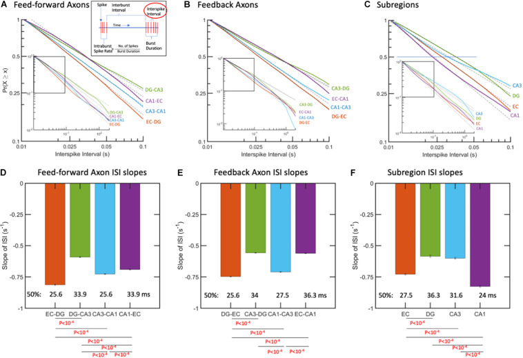
FIGURE 3.
Interspike intervals (ISIs) followed log-log distributions (linear in log-log space). (A) ISIs as complementary cumulative probabilities in feed-forward axons. More spikes at shorter time intervals are from fastest spiking in EC-DG axons. Longer ISIs from slowest spiking axons are in DG-CA3. (B) ISIs in feedback axons. Fastest spiking in axons from DG back to EC. (C) Subregional ISIs are shortest (fastest) in the CA1 subregion. R2 > 0.99 for all fits. N = 10 arrays. (D) Feed-forward axon ISI slopes of which EC-DG is 27% faster than DG-CA3 and each of the others. The median ISI (p = 0.5) for EC-DG from A occurred earliest at 25.6 ms for EC-DG. (E) Feedback axon slopes of ISI distributions with DG back to EC significantly faster than the others and shortest median. (F) Subregion neuron ISI slopes with CA1 fastest. P-values calculated using ANCOVA followed by Tukey-HSD. The error bars represent the 95% confidence intervals obtained by ANCOVA.

