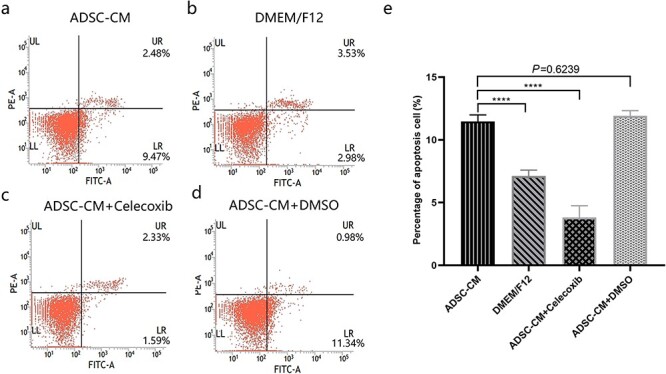
Figure 5.

COX-2 inhibition ameliorated cell apoptosis induced by ADSC-CM. (a, b, c, d) The apoptosis was assessed by Annexin V FITC and phycoerythrin staining in mesangial cells by flow cytometric analysis. (e) The percentage of apoptotic cells in keloid dermis-derived fibroblasts cultured in ADSC-CM with or without COX-2 inhibition, ****p < 0.0001 vs ADSC-CM group, n = 4. ADSC-CM conditioned medium of adipose-derived stem cells, COX-2 cyclooxygenase-2, DMEM Dulbecco’s modified Eagle’s medium, DMSO dimethyl sulfoxide, F12 Ham’s F-12, Bcl-2 B-cell lymphoma-2, LR viable apoptotic cells, UR non-viable apoptotic cells, UL debris and damaged cells; LL normal cells; FITC fluorescein isothiocyanate
