Figure 1.
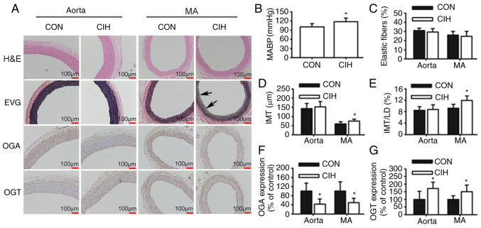
CIH elevates MABP and induces changes in vascular morphology and protein expression. (A) Representative H&E and EVG staining of vessel walls. Immunohistochemical staining for OGA and OGT. Arrowheads indicate disrupted elastic lamella. (B) MABP was increased by CIH. (C) No differences in the content of elastic fibers were observed. (D and E) Changes in vascular wall thickness. Changes in the protein expression levels of (F) OGA and (G) OGT. Scale bar, 100 µm. The groups were as follows: i) CON, normoxia (21% O2); and ii) CIH, intermittent hypoxia cycles (6–8% O2 for 2 min and 21% O2 for another 2 min). Data are presented as the mean ± SD (n=5-20). *P<0.05 between groups. CIH, chronic intermittent hypoxia; MABP, mean arterial blood pressure; H&E, hematoxylin and eosin; EVG, elastin Van Gieson; OGA, O-GlcNAcase; OGT, O-GlcNAc transferase; CON, control; MA, mesenteric artery; LD, luminal diameter; IMT, intima-to-media thickness.

