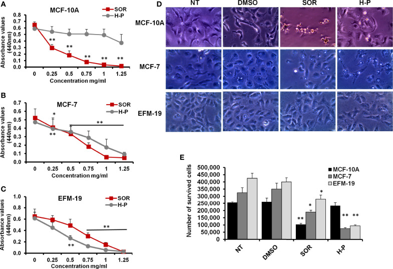
Figure 1.
The cytotoxic effects of SOR and H-P extract in normal human mammary cells and breast cancer cells. (A) The cell viability rate of MCF-10A cells treated with different concentrations of SOR or H-P extract that indicates the CC50 of each one using WST-1 assay, n = 4. (B) The cell viability rate of MCF-7 cells treated with different concentrations of SOR or H-P extract that reveals the CC50 of each inhibitor using WST-1 assay, n = 4. (C) The cell viability rate of EFM-19 cells treated with the same concentrations of SOR or H-P extract and revealed the CC50 of each on other breast cancer cells using WST-1 assay, n = 4. Error bars indicate the standard deviation (SD) of four different replicates. (D) Representative inverted microscopy images of cells morphology two days upon treatment with SOR or H-P extract in comparison with DMSO-treated cells and untreated cells (NT). (E) The number of survived cells treated with either SOR or H-P extract, n = 3. Error bars indicate the SD of three independent experiments. A Student’s two-tailed t-test was used for the significance analysis of represented values. *p ≤ 0.05 and **p ≤ 0.01.

