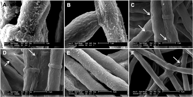
FIGURE 6.

Scanning electron micrographs of the A. alternata hyphae grown in (A) MM, (B) MMT, (C) MMP, (D) MM + pyroquilon, (E) MMT + pyroquilon and (F) MMP + pyroquilon. Arrows indicate holes in the cell wall when the fungus is grown in MM + pyroquilon, MMP and MMP + pyroquilon.
