Figure 1.
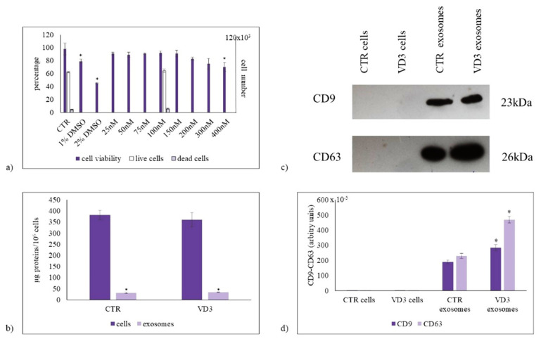
The effect of vitamin D3 on cell viability and exosome purification. (a) Left ordinate, effect of VD3 on HN9.10 cell viability (in gray). Cells were cultured with increasing doses of VD3 from 25 nmol/L to 400 nmol/L for 24 h and the viability was measured by MTT assay. Values were reported as percentage viability of the control sample (CTR). 1% DMSO and 2% DMSO were used as positive controls; right ordinate, live cells (in white) and dead cells (in light violet) under 100 nmol/L VD3 treatment evaluated by trypan blue exclusion assay; (b) Protein content in cells and exosomes; (c) Immunoblotting analysis of CD9 and CD63, as markers of exosome purification. The position of the 23 kDa for CD9, and 26 kDa for CD63 was evaluated in relation to the molecular-weight size markers. (d) The area density was quantified by Chemidoc Imagequant LAS500 by specific IQ programm. (case number:6) CTR, control sample; VD3, vitamin D3 sample. Data were expressed as mean ± SD of three independent experiments performed in duplicate. * p < 0.05 versus the control sample.

