Figure 3.
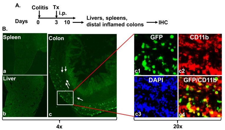
Intraperitoneally administered MAC-CYP cells migrated specifically into inflamed colons. (A) Experimental design. Tx: MAC-CYP-GFP cells. (B) Representative 4× images of MAC-CYP-GFP-treated animals show immunostaining of GFP and CD11b of the spleen (a); liver (b); and distal inflamed colons (c). Arrows indicate GFP+ cells that have migrated to the proximity of crypts. The boxed area is further enlarged (20×) for viewing the staining of GFP (c1), CD11b (c2), and DAPI (c3). (c4) shows GFP and CD11b merged staining.

