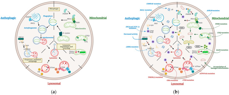
Figure 1.
(a) Schematic of the normal autophagic, mitochondrial, and lysosomal pathways. The tight relationship between these pathways that become altered in PD enables the recycling of deficient organelles and degradation of outer material. Black solid arrows show the normal biological pathway. (b) Schematic of PD disrupted pathways. Dotted arrows indicate an up-regulation of the biological pathway specified due to variants in the genes involved. Meanwhile, inhibiting dotted lines indicate the down-regulation/inhibition of the biological pathway specified due to gene variants. Colored solid arrows show biological pathways that become disrupted in PD. Accumulation of damaged mitochondria and Lewy bodies due to dysfunctional lysosome degradation generates the typical PD physiopathology. Designed with biorender.com.

