Figure 3.
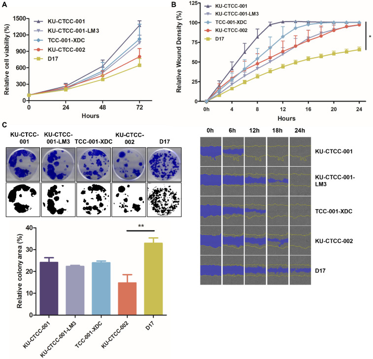
General growth characteristics of TCC cell lines. (A) Comparison of proliferation ability in cell lines using the MTT assay. All canine TCC cell lines proliferated faster than D17 cells (p = 0.2441, two-way repeated measure ANOVA). KU-CTCC-001 proliferated the fastest. There was no significant difference between KU-CTCC-001 and TCC-001-XDC. Error bars represent SEM. (B) The results of the wound-healing assay. All TCC cell lines filled the wound area within 24 h. Error bars represent SD. Statistical significance was determined by one-way ANOVA; * p < 0.05 (C) Representative images of the colony-forming assay. Error bars represent SEM. Statistical significance was determined by one-way ANOVA; ** p < 0.01. All experiments were performed in triplicate.

