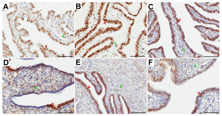
Figure 3.
Representative light micrographs of the nuclear immunolocalization (brown color) of progesterone receptor (PR) in the glandular epithelium (red arrowheads) and the stroma (green arrowheads) of the ampulla (A–C) and isthmus (D–F) of the fallopian tubes in postmenopausal women for whom 1–5 years (A,D), 6–10 years (B,E) and ≥11 years (C,F) had elapsed between the last menstrual period and surgery. Note that in the glandular epithelium, immunonegative PR cells in the form of clusters were usually observed. Scale bar—50 µm.

