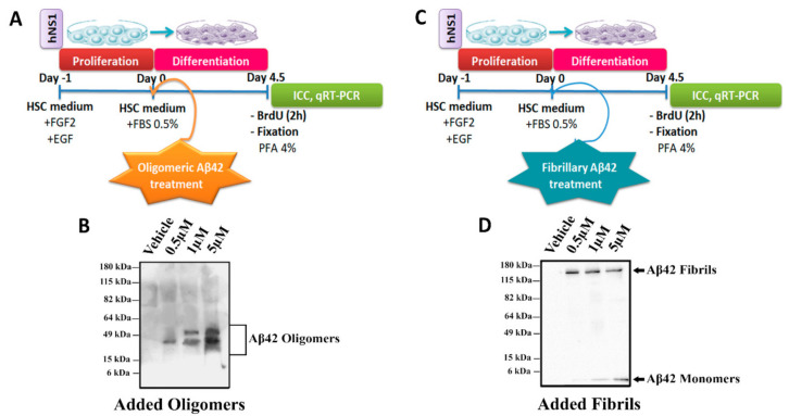
Figure 1.
Schematic representation of the experiments and WB analysis. Schematic view of hNS1 cells’ differentiation protocol (See Materials and Methods section) for (A) Aβ42 oligomers and (C) Aβ42 fibrils. (B) Representative WB analysis of Aβ42 forms (using 4G8 antibody) present in extracellular medium used for oligomeric Aβ42 treatment. (D) Representative WB analysis of Aβ42 forms (using 4G8 antibody) present in extracellular medium used for fibrillary Aβ42 treatment.

