Figure 2.
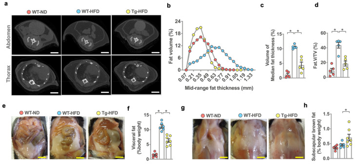
Analysis of adipose tissue of wild-type and miR-29aTg mice upon standard rodent chow (ND) and high-fat diet (HFD) feeding. μCT images of the abdomen fat and subcutaneous fat in the thorax compartment (a); scale bar, 15 mm. miR-29a reversed HFD-induced overall fat thickness (b), median fat thickness (c) and Fat.V/TV (d). Investigations (mean ± S.E.) are calculated from 5 mice. Images of visceral adipose tissue (e); scale bar, 18 mm. miR-29a attenuated HFD-induced visceral fat weight (f). Images of subscapular brown fat tissue (g), scale bar; 18 mm. HFD-fed miR-29a mice showed high subscapular brown fat mass (h). Mean ± S.E. is calculated from 6–7 mice. * p < 0.05 upon ANOVA test and Bonferroni post hoc test. ND, standard rodent chow; HFD, high-fat diet.

