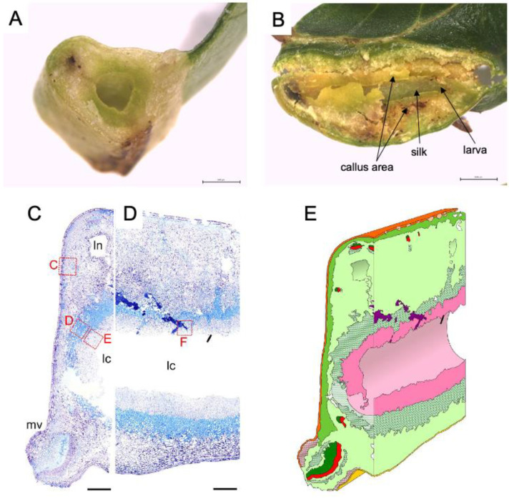
Figure 2.
Inside of leaf gall on Glochidion obovatum. (A) Transverse section. (B) Sagittal section showing callus-like cells, silk covering the chamber wall, and larva inside the chamber. (C,D) Histological transverse (C) and sagittal (D) sections. Ln: lacuna, lc: larva chamber, mv: midvein. (E) 3D representation of gall inside based on histology sections. Dark green: xylem, red: phloem, pink: nutritive tissue, green cross: sclerenchyma, red cross: collenchyma, purple: frass. Bars: A, B, 1 mm; C, D, 0.5 mm. Panels are cited from [7,18].

