
Figure 1.
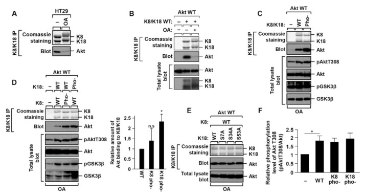
K8/K18–Akt interaction is downregulated by K18 S7/34/53 phosphorylation. (A) HT29 cells were treated with okadaic acid (OA, 1 µM) for 2 h, and then Akt interaction was tested by immunoprecipitation against K8/K18 followed by immunoblotting with Akt antibody. (B) BHK-21 cells were transfected with Akt WT and K8/K18 WT, and treated with OA (0.5 µM) for 2 h. K8/K18 immunoprecipitates and total lysates were prepared and immunoblotted with the indicated antibodies. (C) K8/K18 WT or phosphorylation-deficient mutants (pho-) were transfected into BHK-21 cells together with Akt WT, and then OA was treated 2 h before harvesting cells. K8/K18–Akt interaction was tested by coimmunoprecipitation assay, and cell lysates were immunoblotted with phosphorylated Akt and phosphorylated GSK3 antibodies. K8/K18 pho- indicates K8 S21/22/24/37/43/74/432/451A and K18 S7/34/53A. (D) BHK-21 cells were transfected with the indicated keratin constructs together with Akt WT. Immunoprecipitation was performed with the cells after a 2-h treatment of OA. Each keratin construct is represented as follows: K8 pho-, K8 S21/22/24/37/43/74/432/451A; K18 pho-, K18 S7/34/53A. The graph represents the means ± S.E. of three independent experiments. * indicates p < 0.05. ‘n.s.’ indicates ‘not significant’. (E) Akt WT and K8 WT were transfected into BHK-21 cells with the K18 single-site phosphorylation-deficient mutant (K18 S7A, K18 S34A, or K18 S53A). The cells were treated with OA for 2 h, and the Akt interaction with K8/K18 was tested by the immunoprecipitation assay. (F) The transfected BHK-21 cells were prepared as described in panel D. Akt activity was determined by immunoblotting cell lysates with antibody against phosphorylated Akt T308. The data were obtained from three independent experiments, and the mean values ± S.E. are shown in the graph. * indicates p < 0.05.