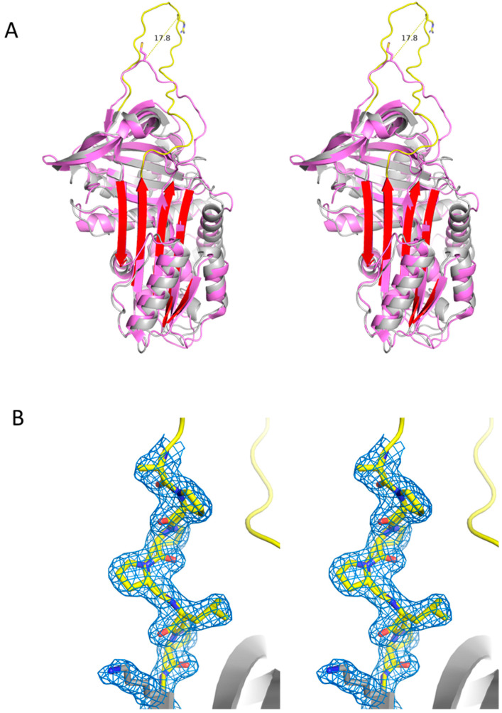
Figure 5.
Crystal structure of native Iripin-8. (A) Stereo view of a ribbon diagram of Iripin-8 (gray with yellow RCL and red beta sheet A) superimposed with alpha-1-antitrypsin (PDB code 3ne4). The P1 side chains of both molecules are represented as sticks, and the distance between their Cα atoms is shown. (B) Stereo view of a close-up of the P′ region with surrounding electron density (contoured at 1 times the RMSD of the map), forming a rigid type II polyproline helix.

