Figure 4.
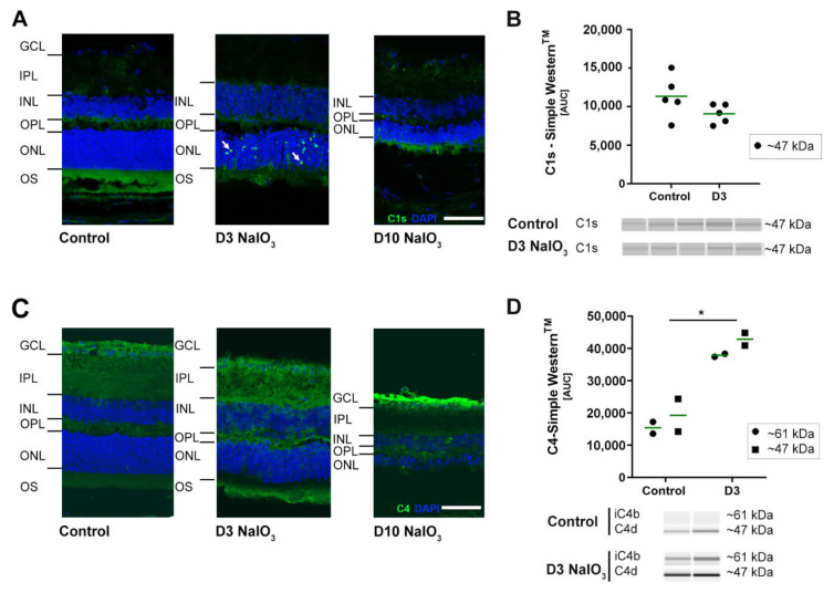
Classical pathway proteins C1s and C4 accumulated in NaIO3 damaged the retinas. (A) C1s immunoreactivity (green) was localised (white arrows) to the ONL at three days after the NaIO3 treatment. At ten days, no specific C1s staining remained visible. DAPI staining (blue) delineated cell nuclei. (B) C1s protein levels were not changed in the Simple WesternTM three days after the NaIO3 treatment. (C) C4 (green) was observed in the nerve fibre layer (NFL) ten days following the NaIO3 treatment. In contrast, no distinct C4 staining was visible at day 3. Scale bar: 50 µm. (D) C4 cleavage products C4d (47 kDa) and iC4b (61 kDa) were increased at day 3 after the NaIO3 treatment in all samples compared to the untreated control using Simple WesternTM technology. (B,D) Intensity of the signals was measured using ImageJ software. * 0.01 < p < 0.05 (one-way ANOVA and Tukey’s multiple comparisons test). The full blots are shown in Supplementary Figure S3C,D.

