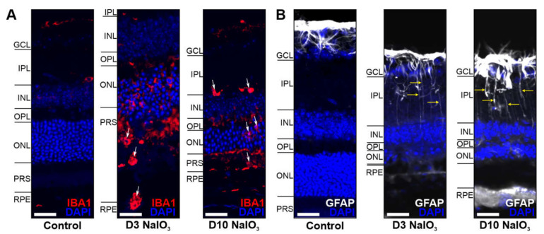
Figure 7.
Glia cells were activated in the NaIO3-damaged retina. (A) Microglia/monocytes (arrows) migrated into the photoreceptor outer segment layer at day 3 following the NaIO3 treatment and were localised in the IPL at day 10. (B) GFAP (white), a marker for activated Müller cells, was enhanced in the GCL after NaIO3 treatment during the investigated period. Scale bar: 20 µm.

