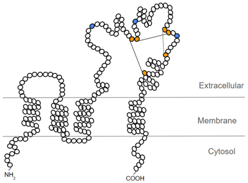
Figure 2.
Schematic representation of the probable structure of CD63. In orange, the cysteine residues that form the 3 disulfide bridges are depicted. In blue, the N-glycosylation sites present in the large extracellular loop of CD63 are represented. Figure based on the representation proposed by Warner et al. [28].

