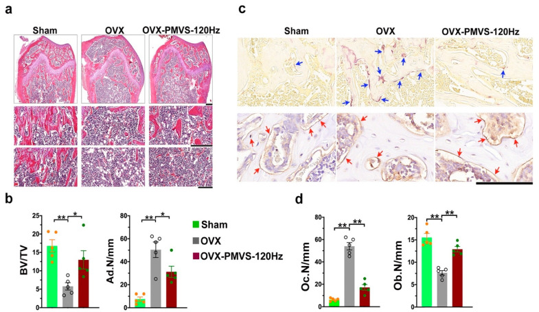
Figure 4.
Histological analysis of trabecular bone morphology osteoclasts and osteoblasts in distal femoral bone specimens. Hematoxylin-eosin staining images revealing mild loss in trabecular bone loss (scale bar, 200 µm in upper panels; scale bar, 50 µm in middle panels) and moderate marrow adipocyte formation (scale bar, 50 µm in lower panels) in OVX bone tissue upon PMVS intervention (a). PMVS intervention reversed OVX-induced BV/TV loss and Ad.N upregulation (b). Tartrate-resistant acid phosphatase histochemical staining images (blue arrows) showing few osteoclasts and osteocalcin immunohistochemical staining (red arrows) images showing plenty of osteoblasts in OVX bone tissue upon PMVS intervention (c); scale bars, 10 µm. PMVS intervention attenuated OVX-induced osteoclast formation and osteoblast loss (d). Difference of investigations (mean ± S.E.; n = 4–5) were analyzed using Dunn’s nonparametric comparison test, where asterisks * and ** resemble p < 0.05 and p < 0.01, respectively.

