Figure 5.
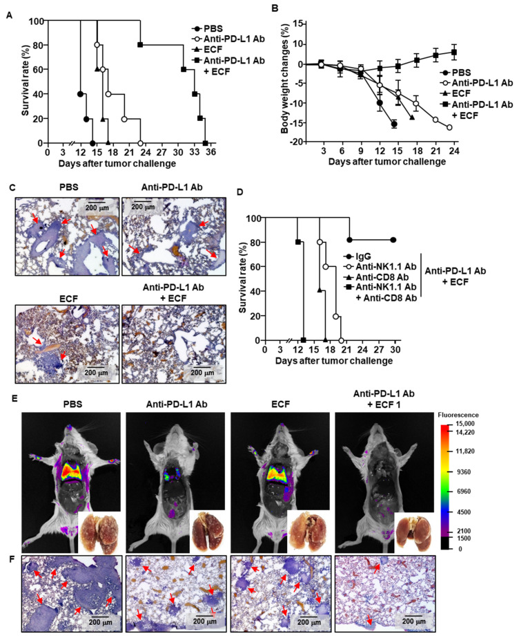
ECF enhances anti-PD-L1 antibody-induced anti-cancer immunity. C57BL/6 mice were challenged intravenously (i.v.) with 0.5 × 10⁶ B16 cells. As indicated in the materials and method, the mice were treated with PBS 10 mg/kg of anti-PD-L1 antibody (Ab), 50 mg/kg of ECF and the combination of ECF and anti-PD-L1 Ab. (A) The graph showed survival rate of treated mice (n = 5 for each group). (B) Changes of body weight during treatment were shown. (C) Hematoxylin and eosin (H&E) staining of lung showed B16 melanoma cell infiltration, as analyzed on day 10 after tumor injection. Red arrows indicated infiltrated tumor cells. (D) Survival rate, as measured in natural killer (NK)- or CD8 T cell-depleted mice (n = 5 for each group). (E,F) BALB/c mice were injected i.v. with 0.5 × 10⁶ CT-26-iRFP cells. The mice received with PBS, 10 mg/kg of anti-PD-L1 Ab, 50 mg/kg of ECF or the combination of anti-PD-L1 Ab and ECF for a 3 day-interval. (E) Fluorescence imaging of iRFP in the mice on day 14 after CT-26-iRFP cell administration in BLAB/c mice, n = 5. (F) CT-26 cell infiltration in lung was analyzed by H&E staining of lung tissue 14 days after CT-26 injection in BALB/c mice. Red arrows indicated infiltrated tumor cells.

