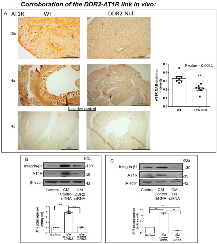
Figure 6.
Corroboration of the DDR2-AT1R link in vivo. (A) Representative image showing 3,3′-diaminobenzidine (DAB) staining of AT1R protein in myocardial tissue sections of 10-week-old WT and DDR2-null mice. ** p < 0.01 versus WT (n = 7). (B) Sub-confluent quiescent cultures of H9c2 cells were treated for 24 h with conditioned medium (CM) derived from control siRNA-treated cardiac fibroblasts or DDR2-silenced cardiac fibroblasts. Quiescent cultures of H9c2 in M199 without serum were used as the control for basal AT1R protein expression in these cells. AT1R protein expression in H9c2 cells was examined by Western blot analysis and normalized to β-actin. ** p < 0.01 (comparisons as depicted in the Figure), (n = 3, for the conditioned medium experiments, cardiac fibroblasts were from three isolations from three rats). (C) Sub-confluent quiescent cultures of H9c2 cells were treated for 24 h with conditioned medium (CM) derived from control siRNA-treated cardiac fibroblasts or fibronectin (FN)-silenced cardiac fibroblasts. Quiescent cultures of H9c2 in M199 without serum were used as the control for basal AT1R protein expression in H9c2 cells. AT1R protein expression was examined by Western blot analysis and normalized to β-actin. ** p < 0.01 (comparisons as depicted in the Figure), (n = 3, for the conditioned medium experiments, cardiac fibroblasts were from three isolations from three rats).

