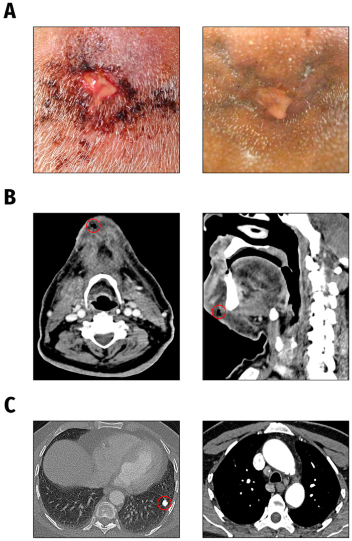
Figure 4.
Clinical presentation. (A). Submental wound findings at initial presentation (left) and following antibiotic treatment one month later (right). (B). CT head and neck findings: subcutaneous air accumulation and circumscribed fluid in the chin region. (C). CT chest scan findings: calcified focal lesion in the left lung as well as mediastinal and pulmonary lymphadenopathy.

