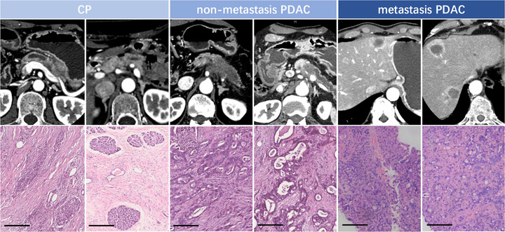
FIGURE 1.

Representative imaging features and pathological information of patients with CP, nonmetastasis PDAC, and metastasis PDAC. Representative imaging results of the patients were performed using enhanced CT. Hematoxylin and eosin staining results of tumor tissue specimens derived from patients are shown. Magnification is ×200. Scale bar, 200 μm
