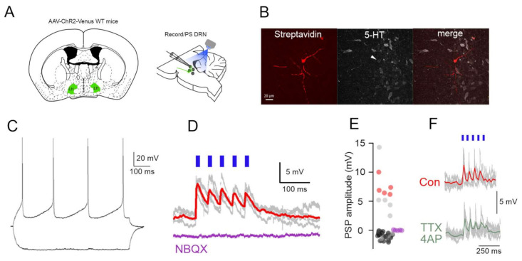
Figure 2.
Functional excitatory projections from LH to DRN. (A) Schematics of the experimental design for the in vitro circuit mapping experiments. (B) Fluorescent images (Streptavidin, 5-HT immunostaining, merged) of a DRN 5-HT neuron responding to LH axonal photostimulation. (C) Membrane potential responses of the neuron in (B) to hyperpolarizing and depolarizing current steps. (D) The photostimulation of LH axons leads to EPSPs in the neuron shown in (B,C) (gray traces: individual voltage responses, red trace: average) that are blocked by the iGluR antagonist (NBQX, purple trace). (E) Distribution of PSP amplitudes in all the recorded neurons (n = 51) (red circles indicate identified DR-5-HT neurons, gray circles unidentified neurons, purple circles EPSP amplitudes following NBQX application (n = 5). (F) Persistence of the LH axonal photostimulation induced EPSPs in TTX/4AP in a DRN neuron.

