Figure 1.
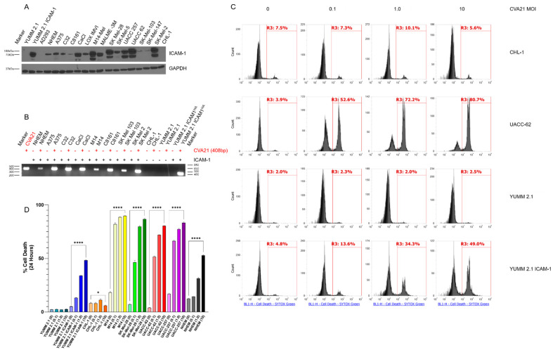
Melanoma cells are susceptible to CVA21 infection and cell death. (A) Immunoblot of a panel of melanoma cell lines probing for ICAM-1. GAPDH was used as a loading control. Mouse YUMM 2.1 cells and lentivirally transduced YUMM 2.1 ICAM-1 cells were used as negative and positive controls for ICAM-1, respectively. (B) RT-PCR of RNA extracted from a panel of melanoma cell lines infected with CVA21 compared to uninfected parental lines. RT-PCR on purified CVA21 stock was used as a positive control for comparison. The RT-PCR produces an amplicon from the 3′ end of the viral genome in the 3Dpol. (C) Assessment of cell death following CVA21 infection at MOI 0, 0.1, 1.0, and 10 at 24 h by flow cytometry in representative cell lines. SYTOX Green was used to stain dead cells. (D) Quantitation of cell death from the flow cytometry assay represented in panel (C). Samples were run in triplicate and analyzed using a one-way ANOVA test for statistical differences. Error bars represent the standard deviation between replicates. p values < 0.05 were considered significant (p < 0.05 *, < 0.0001 ****).

