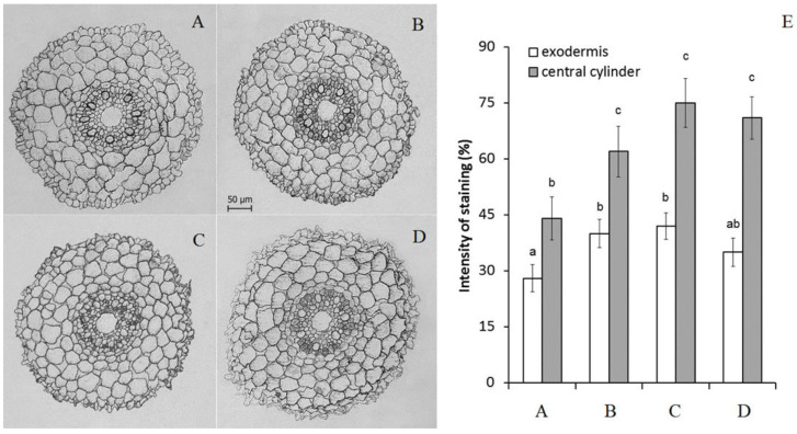
Figure 2.
Immunolocalization of ABA (A–D) and intensity of staining for ABA of exodermis and central cylinder (E) (means ± SE, arbitrary units, maximal staining taken as 100%, minimal as 0%) in root sections untreated (A), treated with 10 μM ABA (B) and its combination with 10 μM diphenylene iodonium chloride (C) and 10 mM ascorbic acid (D). Data represent the mean ± S.E. (n = 50) of arbitrary units; maximal staining was taken for 100%, and minimal staining was 0%. Significantly different means for each variable are labelled with different letters (p ≤ 0.05, LSD test).

