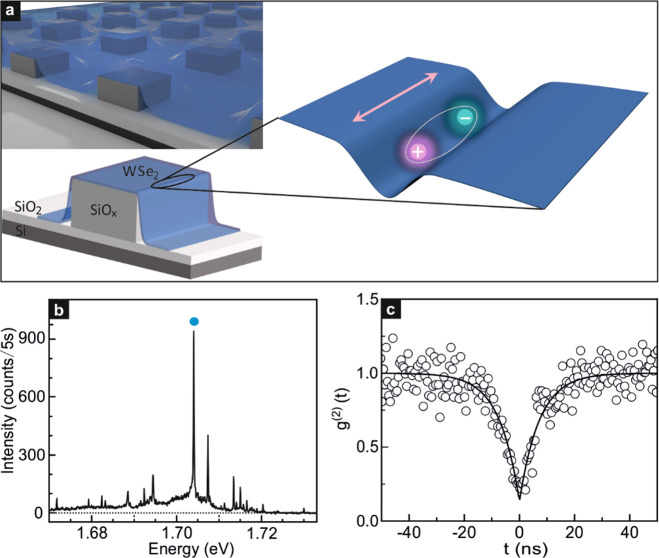
Figure 1.
Sample geometry for generating quasi-1D localized excitons. (a) Schematic cross section of a WSe2 monolayer placed on top of an array of SiOx pillars (left) and schematic illustration of a quasi-1D localized exciton trapped by the strain-induced potential at the edge of the SiOx pillar upon laser illumination (right). The arrow marks the orientation of the oscillating exciton dipole. It is parallel to the one-dimensional strain-induced potential along the pillar edge. (b) Micro-photoluminescence (μ-PL) spectrum obtained when illuminating the WSe2 monolayer at the edge of SiOx pillar with 633 nm laser. The blue dot highlights the emission peak at 1.704 eV investigated in this Letter. It stems from localized bright excitons. The experimental data were recorded at approximately 2 K. (c) Second-order photon-correlation measurement of another localized exciton emission peak, measured near 4 K when pumping with a CW laser with a wavelength at 658 nm. The black solid line is a fit to the data. At zero time delay, g(2)(0) = 0.13 ± 0.04.

